




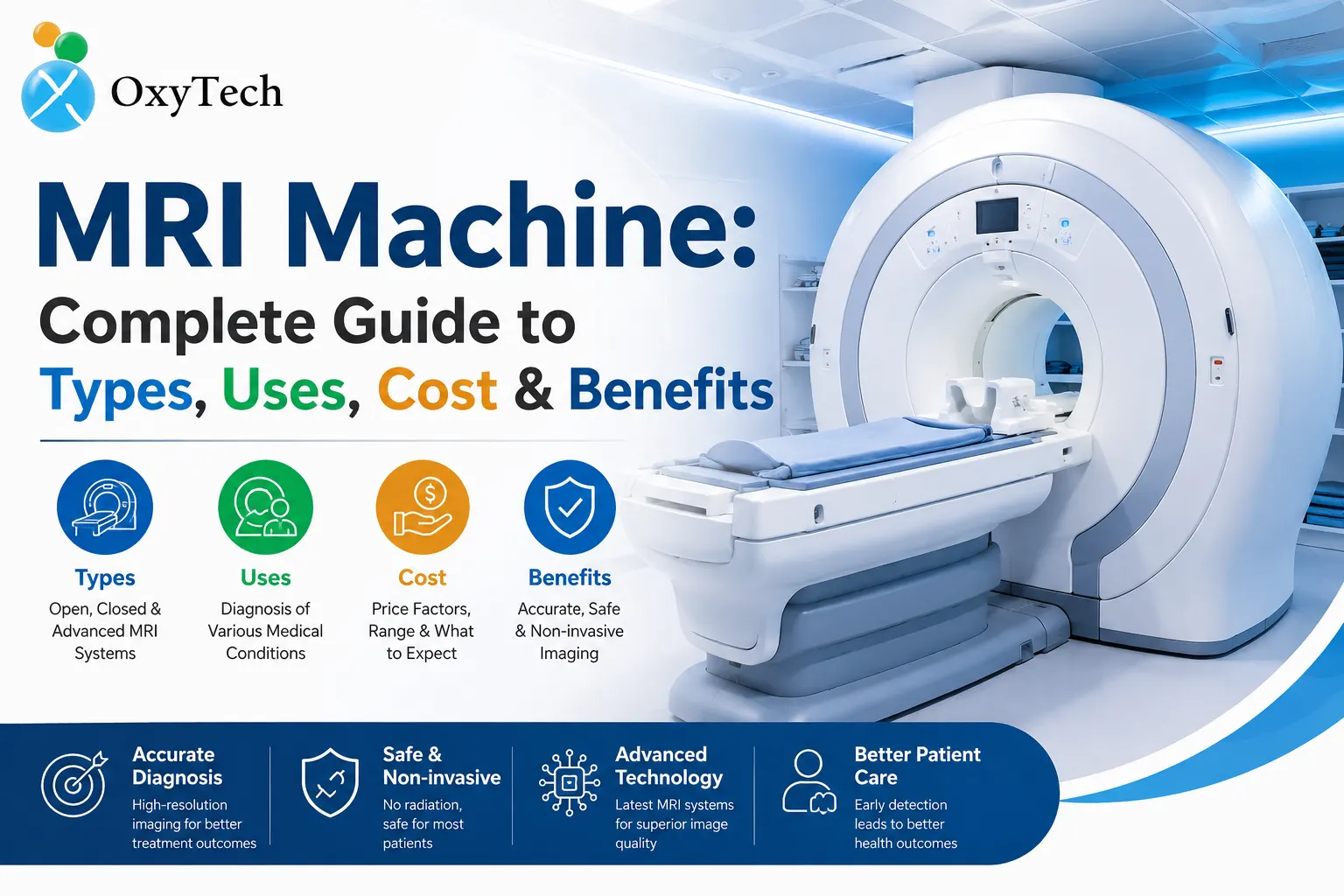
MRI Machine: Complete Guide to Types, Uses, Cost & Benefits
Magnetic Resonance Imaging (MRI) is one of the most powerful, noninvasive diagnostic tools in modern medicine. Unlike X-rays or CT scans, MRI uses magnetic fields and radiofrequency pulses to produce detailed images of soft tissues, organs, and structures inside the body. This guide explains how MRI machines work, the main types of MRI systems, clinical and nonclinical uses, typical costs, risks and benefits, and practical information patients and healthcare buyers should know. The article emphasizes evidence-based facts, clear explanations, and actionable tips to help clinicians, patients, and facility decision-makers make informed choices.
How MRI Works — Simple, Accurate Explanation
- Basic physics: MRI detects signals from hydrogen nuclei (protons) in water and fat. When placed in a strong magnetic field, protons align with that field. Radiofrequency pulses briefly disturb that alignment; when protons relax back, they emit signals captured by receiver coils and converted into images.
- Image contrast: Differences in tissue composition and relaxation times (T1, T2) create contrast between structures, allowing radiologists to distinguish gray matter, white matter, tumors, edema, and more.
- Safety and no ionizing radiation: MRI does not use ionizing radiation (unlike X-ray/CT), making it safer for repetitive imaging when clinically indicated.
- Components: Main magnet (creates field strength, measured in tesla, T), gradient coils (spatial encoding), RF coils (transmit/receive), and computer/software (image reconstruction and postprocessing).
Types of MRI Machines
- Closed-bore MRI (High-field MRI)
- Field strength: typically 1.5T and 3T for clinical use.
- Advantages: high image resolution, faster scans, excellent for neuro, musculoskeletal, cardiovascular, and body imaging.
- Disadvantages: longer tunnel, potential claustrophobia, stronger magnetic field imposes stricter device/implant limits.
- Open MRI
- Geometry: C-shaped or wide-open design that reduces enclosure and claustrophobia.
- Field strength: usually 0.2–1.0T (low- to mid-field).
- Advantages: comfort for claustrophobic or larger patients, easier positioning for pediatrics and interventional guidance.
- Disadvantages: lower image resolution; some exams may be less diagnostic than high-field MRI.
- Wide-bore MRI
- Hybrid: provides better patient comfort than closed bore while maintaining high-field strength (commonly 1.5T).
- Advantages: balance of image quality and comfort; common in many modern imaging centers.
- Extremity MRI
- Dedicated for arms, legs, hands, feet, knees, shoulders.
- Advantages: targeted scans, lower cost, comfortable positioning; often open design.
- Disadvantages: not suitable for head, chest, or abdominal imaging.
- Ultra-high-field MRI (7T and above)
- Research and specialized clinical use: exceptional resolution for neuroimaging and advanced research.
- Advantages: superior detail for small structures.
- Disadvantages: cost, infrastructure (shielding), safety/implant considerations; not widely available for routine clinical imaging.
- Portable and point-of-care MRI
- Low-field, compact systems designed for bedside use, emergency departments, or field medicine.
- Advantages: immediate imaging where moving the patient is risky.
- Disadvantages: lower resolution; suitable for triage or specific indications rather than full diagnostic workups.
Clinical Uses of MRI
- Neurology and brain imaging
- Indications: stroke evaluation (with diffusion-weighted imaging), brain tumors, multiple sclerosis, epilepsy focus localization, dementia workup, traumatic brain injury.
- Key sequences: T1, T2, FLAIR, diffusion-weighted imaging (DWI), susceptibility-weighted imaging (SWI), contrast-enhanced sequences.
- Spine and spinal cord
- Indications: disc herniation, spinal stenosis, infection, neoplasm, spinal cord compression, demyelinating diseases.
- Strengths: multiplanar imaging, excellent soft-tissue contrast.
- Musculoskeletal (MSK)
- Indications: ligament/tendon injuries, cartilage defects, bone marrow edema, sports injuries, joint pathology.
- MRI advantage: sensitive to soft tissue and early bone changes without radiation.
- Cardiovascular MRI (Cardiac MRI)
- Indications: cardiomyopathies, myocardial viability/scar, congenital heart disease, cardiac masses, aortic disease.
- Techniques: cine imaging for heart function, late gadolinium enhancement for scar/fibrosis.
- Abdominal and pelvic MRI
- Indications: liver lesions, pancreatic disease, pelvic masses, prostate imaging (multi-parametric MRI for prostate cancer), inflammatory bowel disease evaluation.
- Breast MRI
- Indications: screening for high-risk patients, problem solving after abnormal mammogram/ultrasound, preoperative staging.
- Advantage: high sensitivity for invasive cancer and multifocal disease.
- Oncologic imaging and staging
- MRI used for local staging, treatment planning, and follow-up for many cancers (brain, liver, prostate, gynecologic, musculoskeletal).
- Pediatrics and fetal MRI
- Applications: congenital anomalies, brain development, spine abnormalities; fetal MRI complements ultrasound for complex cases.
- Functional MRI (fMRI) and advanced techniques
- fMRI: maps brain activity by detecting blood-oxygen-level-dependent (BOLD) signal; used in neurosurgical planning and cognitive research.
- Diffusion tensor imaging (DTI): maps white-matter tracts, useful in pre-surgical planning and assessing traumatic axonal injury.
- MR spectroscopy: measures tissue metabolites to help characterize tumors and metabolic disorders.
- Perfusion imaging: measures blood flow to detect ischemia or tumor vascularity.
Benefits of MRI
- Superior soft-tissue contrast: best among common imaging modalities for differentiating soft tissues.
- No ionizing radiation: safer for repeated imaging and younger patients.
- Versatility: wide range of sequences tailored to specific clinical questions.
- Multiplanar capability: can produce axial, coronal, sagittal, and oblique images without repositioning the patient.
- Functional and metabolic information: advanced techniques provide physiological and biochemical data beyond anatomy.
Limitations and Risks
- Claustrophobia and patient comfort: closed strong-field systems can be uncomfortable; pre-scan anxiolytics or open/wide-bore MRI can help.
- Contraindications and implant safety: certain metallic implants, pacemakers, cochlear implants, and some aneurysm clips may be unsafe or require specialized MRI‑conditional devices and protocols.
- Gadolinium-based contrast agents (GBCA): generally safe, but small risk of allergic reactions and potential gadolinium retention in tissues; use lowest effective dose and consider macrocyclic agents when contrast is needed.
- Pregnancy: MRI without contrast is generally considered safe after the first trimester when clinically necessary; avoid gadolinium unless critical.
- Clauses on artifacts and limitations: motion, metal hardware, and some implants cause artifacts that reduce diagnostic quality.
- Cost and availability: MRI exams are more expensive and less available in low-resource settings compared with X-ray/ultrasound.
MRI Cost: What to Expect
- Factors that influence cost:
- Type of MRI scan: brain, spine, cardiac, or whole-body—complexity and sequences determine time and cost.
- Use of contrast: contrast-enhanced studies increase cost.
- Field strength and facility: high-field 3T scans and advanced centers charge more.
- Location and healthcare system: prices vary widely by country, public vs private hospitals, and outpatient imaging centers.
- Rough price ranges (indicative; vary by market and year)
- Low-cost markets or subsidized public hospitals: USD 50–200 for basic regional MRI.
- Typical private outpatient centers (India context reference): INR 3,000–12,000 for single-region MRI, INR 10,000–30,000 for advanced or contrast studies (approximate; vary by city and facility).
- High-income countries: USD 400–3,000+ depending on exam, contrast use, and facility.
- Advanced or specialized MRI (cardiac MRI, 3T, functional studies): significantly higher.
- Ownership and operational costs for facilities:
- Purchase price: new MRI systems range roughly from USD 500,000 for low-field or refurbished units up to USD 3 million+ for high-end 3T systems (installation and site prep add substantial cost).
- Installation: shielding, cooling systems (cryogens for superconducting magnets), room modifications, and power upgrades.
- Maintenance and service: annual service contracts often 8–15% of purchase price; coil replacements and software upgrades add cost.
- Staffing: trained MRI technologists, radiologists, and safety oversight.
- Cost-saving strategies for patients and buyers:
- Shop around: compare prices at multiple imaging centers.
- Use public or teaching hospitals for lower-cost options.
- Consider whether contrast or high-field imaging materially changes the clinical decision.
- For facilities: refurbished units, service contracts negotiation, and sharing resources across hospitals.
Choosing the Right MRI Exam
- Match the clinical question: order the MRI tailored to the suspected diagnosis (e.g., knee MRI for persistent joint pain, brain MRI with DWI for suspected stroke).
- Field strength considerations: 1.5T is sufficient for most clinical exams; 3T improves resolution for brain, prostate, and MSK but may increase artifacts in some cases.
- With or without contrast: use gadolinium when needed to differentiate lesions, detect breakdown of the blood-brain barrier, or evaluate tumor vascularity; otherwise noncontrast protocols often suffice.
- Accessibility and patient factors: for claustrophobic or larger patients, prefer wide-bore or open MRI; for pediatric patients, sedation pathways and child-friendly centers improve success.
- Implant and device verification: verify MRI compatibility (MRI-conditional) and follow manufacturer instructions on scanning conditions.
Preparing for an MRI: Practical Tips for Patients
- Safety screening: disclose implants, pacemakers, metal fragments, tattoos, or recent surgeries.
- Clothing and personal items: remove jewelry, watches, credit cards, hairpins, and metallic objects.
- Eating and medications: usually no special food restrictions unless sedation or contrast is planned.
- Claustrophobia: request a wide-bore or open MRI, use relaxation techniques, or ask about light sedation.
- Communication during scan: technologists can talk and provide an alarm button; follow breathing instructions for certain sequences.
- After contrast: drink fluids to help eliminate gadolinium; report allergies or delayed reactions.
Interpretation and Reporting
- Radiologist’s role: analyze sequences, correlate with clinical history, and provide a structured report with findings, impressions, and recommendations.
- Structured reporting: improves clarity—clinical history, technique, comparison studies, findings, and diagnostic impression.
- Follow-up recommendations: when appropriate, suggest additional imaging, biopsy, or referral to specialists.
Emerging Trends and Innovations
- Artificial intelligence (AI) in MRI
- Applications: image reconstruction to speed scans, automated lesion detection/segmentation, workflow optimization, and quantitative imaging biomarkers.
- Benefits: shorter scan times, improved throughput, enhanced diagnostic consistency.
- Accelerated imaging techniques
- Compressed sensing, parallel imaging, and deep-learning reconstruction reduce acquisition time and motion artifacts.
- Hybrid imaging
- PET/MRI combines metabolic information with high soft-tissue contrast—valuable in oncology and neurology.
- Low-field MRI resurgence
- New low-field systems use advanced reconstruction to provide useful clinical images at lower cost and with fewer infrastructure demands—improving access in resource-limited settings.
- MR-guided interventions
- Real-time MRI guidance for targeted biopsies, focused ultrasound therapies, and minimally invasive procedures.
Patient Safety and Regulatory Considerations
- Quality standards: accreditation (e.g., ACR in the U.S.) ensures safety protocols, image quality, and staff training.
- MR safety officers and policies: facilities should maintain MR safety committees, screening protocols, and emergency procedures.
- Gadolinium use: follow guidelines to minimize risk—prefer macrocyclic agents for patients at higher risk and document cumulative exposure.
Common Patient Questions (FAQs)
- Is MRI painful? No; it’s painless, though some sequences can feel noisy. Ear protection and music reduce noise and discomfort.
- How long does an MRI take? Typical regional MRI: 20–45 minutes. Complex or contrast studies: up to 60–90 minutes.
- Can I have an MRI with a pacemaker? Many older pacemakers are contraindicated; newer MRI-conditional devices may allow scanning under specific protocols—check device details and vendor guidance.
- Are MRIs safe during pregnancy? Noncontrast MRI is used when clinically necessary; gadolinium is avoided unless essential.
- Will insurance cover MRI? Coverage depends on clinical indication and insurance policies; preauthorization may be required.
Buying an MRI for a Facility: Checklist
- Clinical needs assessment: expected patient volume, types of exams (neuro, MSK, cardiac, body).
- Field strength choice: 1.5T for general use; 3T for high-resolution and specific advanced applications.
- Site requirements: room size, shielding, cooling (cryogens or dry magnet options), power supply, and HVAC.
- Budget and financing: purchase vs lease, refurbished options, long-term service contracts.
- Vendor support and upgrades: warranty terms, remote diagnostics, software upgrades, and coil availability.
- Staff and training: MRI technologists, physicist support, and radiologist subspecialty expertise.
- Accreditation and compliance: local regulatory approvals, patient safety plans, and quality assurance protocols.
Summary: When to Choose MRI
- Opt for MRI when high soft-tissue contrast, multiplanar imaging, or functional/metabolic information is necessary for diagnosis or treatment planning.
- Consider CT, ultrasound, or X-ray when MRI is contraindicated, unavailable, or when those modalities better address specific clinical questions (e.g., acute bone fractures, lung pathology, or when cost/time constraints apply).
Conclusion
MRI remains indispensable for modern diagnostics, offering unmatched soft-tissue contrast and a growing set of functional tools. Selecting the right MRI type, optimizing protocols, and following safety guidelines ensure accurate, efficient, and patient-centered imaging. Whether you are a patient preparing for a scan, a clinician ordering an exam, or a facility evaluating purchase options, understanding MRI’s capabilities, costs, and limitations helps you get the best outcomes.